38 label the replication fork
M7 DNA Replication Fork Homework.pdf - Name: PCB3063... Label the prokaryotic replication fork with all molecules and structures involved in their correct positions. Include, Gyrase, Helicase, SSBP, DNA Pol I, DNA Pol III, Sliding Clamp, Primase, RNA primer, Okazaki fragment, Ligase, 3' & 5' ends of parental and daughter strands, and indicate overall direction of replication. M7 DNA Replication Fork Homework.docx - Name: PCB3063... Label the Replication Fork in question 1. Take a clear picture of your replication fork and paste it into this word.doc.Type in your answers to questions 2-10. Type in your name above. Save and upload to Canvas. 1. Label the prokaryotic replication fork with all molecules and structures involved in their correct positions.
PDF Bio 102 Practice Problems Chromosomes and DNA Replication 1. Below is a replication fork (one side of an origin): a double-stranded DNA partially opened up to provide single-stranded regions where replication can occur. Draw and label the following: RNA primers (label 3′ and 5′ ends), leading-strand DNA polymerase and new DNA (label 3′ and 5′ ends and show direction of synthesis
Label the replication fork
Draw a labelled diagram of replicating fork. - Toppr Ask DNA replicates in a semi-conservative manner in which each individual strand is copied to form a new molecule of DNA. The two strands can be labelled with isotopes using substrates that contain either normal 1 4 N or its heavy isotope 1 5 N.In an experiment, one strand of DNA was labelled with 1 4 N and the other with 1 5 N (hybrid DNA). The hybrid DNA was then allowed to replicate in the ... DNA Replication - Labeling worksheet with image - Name - StuDocu DNA Replication - Labeling (with word bank) DNA polymerase 3' 5' DNA Ligase Okazaki fragment DNA Primase ... ____ DNA polymerase adds nucleotides in the 5' to 3' direction ____ Replication fork is formed ____ DNA polymerase attaches to the primer ____ Okazaki fragments are bound together by ligase ____ DNA helicase unwinds DNA. DNA Replication Labeling Diagram | Quizlet An enzyme that creates a short RNA primer for initiation of DNA replication. RNA Primer short segment of RNA used to initiate synthesis of a new strand of DNA during replication The primer synthesized by primase enzyme DNA Polymerase on Leading Strand synthesizes new DNA only in the 5' to 3' direction Ligase
Label the replication fork. PDF 12.3 DNA Replication - rvrhs.com Label the other as Eukaryotic DNA. 3. Label both drawings with the following terms: unreplicated DNA, replication fork, origin of replication. Answer the questions. Circle the correct answers. 4. In which type of cell is DNA circular? prokaryotic eukaryotic 5. In which type of cell does replication begin at several points? prokaryotic ... DNA Replication Quiz Questions And Answers - ProProfs Quiz Well, here in this quiz, we will ask you some questions related to this topic. Let's see if you can answer them or not! Questions and Answers. 1. What are the four nitrogen bases for DNA? A. Uracil, Thymine, Adenine, and Guanine. B. Cytosine, Thymine, Adenine, and Guanine. DNA Replication and Repair: DNA Replication | SparkNotes The first step in DNA replication is the separation of the two DNA strands that make up the helix that is to be copied. DNA Helicase untwists the helix at locations called replication origins. The replication origin forms a Y shape, and is called a replication fork. The replication fork moves down the DNA strand, usually from an internal ... The Initiation and Completion of DNA Replication in Chromosomes To replicate such a DNA molecule from end to end with a single replication fork moving at a rate of 50 nucleotides per second would require 0.02 × 150 × 10 6 = 3.0 × 10 6 seconds (about 800 hours). As expected, therefore, the autoradiographic experiments just described reveal that many forks are moving simultaneously on each eucaryotic chromosome.
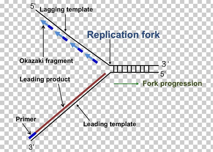
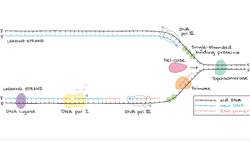
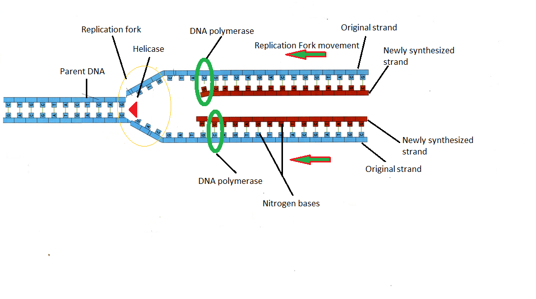
Chapter 6 REPLICATION Flashcards | Quizlet Single-strand binding proteins bind to the single-stranded DNA near the replication fork to keep the fork open. Be prepared to label a replication fork including the leading and lagging strands and Okazaki fragments (in multiple choice format). See Fig. 6.13 on slide 6.3d What is the function of DNA ligase? seals nicks in DNA In the replication fork, label the leading and lagging strands and the ... Label the image... The image shows a replication fork, template DNA strands, and new DNA strands. Label the image by moving the terms to the apropriate targets. Not all terms will be used. RNA primers on the leading and lagging strands Answered: . Draw a replication bubble with both… | bartleby Draw a replication bubble with both replication forks and label the origin of replication, the leading strands, lagging strands, and the 5' and 3' ends of all strands shown in your diagram. Pls help asap! arrow_forward. Draw a molecule of DNA undergoing theta replication. On your drawing, identify (a) origin of replication, (b) polarity (5 ... DNA Replication Fork: Definition & Overview - Study.com The replication fork is the area where the replication of DNA will actually take place. There are two strands of DNA that are exposed once the double helix is opened. One strand is referred to as...
Label The Diagram Showing Dna Replication - Slainie Brousse Label the diagram showing the dna replication. Show dna replication with the help of a diagram only. (a) draw a labelled diagram of a \replicating fork\ showing the polarity. Rna primers (label 3′ and. You should label all the parts of the dna including the covalent and hydrogen bonds. (Get Answer) - Label The Steps Shown For HIV Retroviral Replication ... Label The Steps Shown For HIV Retroviral Replication. (Labels Are Not In Correct Order.) Label the steps shown for HIV retroviral replication. Drag the appropriate labels to their respective targets. Reset Entry by endocytosis Attachment Uncoating CD4 receptor DNA synthesis. Solved 2. Label the Replication Fork in question 1. Draw in - Chegg Label the prokaryotic replication fork with all molecules and structures involved in their correct positions. Include, Gyrase, Helicase, SSBP, DNA Pol I. DNA Pol III, Sliding Clamp, Primase, RNA primer, Okazaki fragment, Ligase, 3 & 5' ends of parental and daughter strands, and indicate overall direction of replication. 2. The E. coli DNA Replication Fork - PubMed DNA replication in Escherichia coli initiates at oriC, the origin of replication and proceeds bidirectionally, resulting in two replication forks that travel in opposite directions from the origin. Here, we focus on events at the replication fork. The replication machinery (or replisome), first assembled on both forks at oriC, contains the DnaB ...
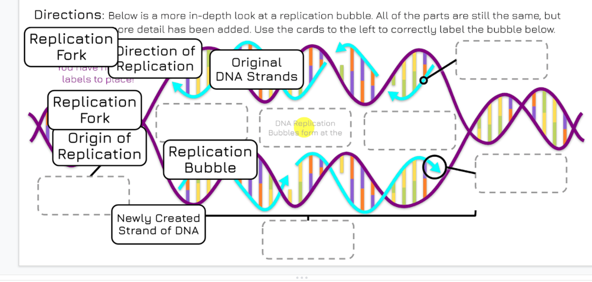
Answered: Directions: Below is a more in-depth… | bartleby
DNA Replication: Review of Enzymes, Replication Bubbles & Leading and ... The fat arrow marks the replication fork. 1. Label the newly made leading strand of DNA, including the 5' and 3' ends. Hint: the leading strand is the one that is made continuously! 2. Label the ...
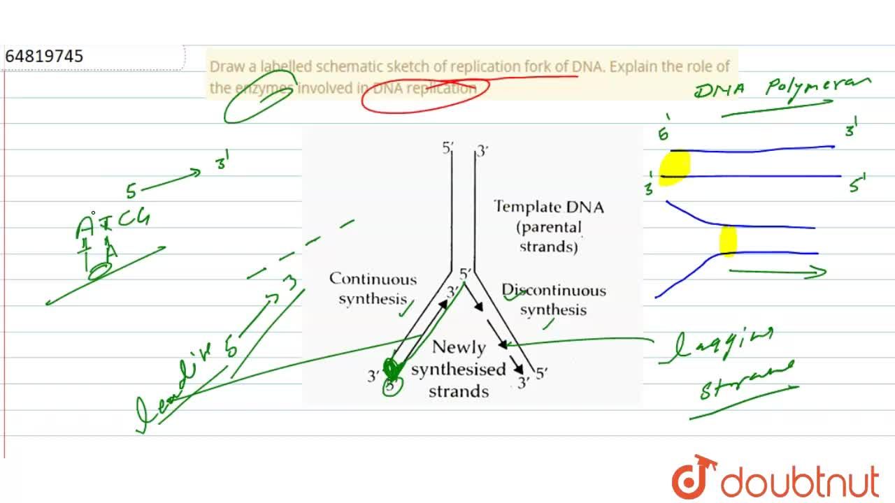
Draw a labelled schematic sketch of replication fork of DNA ...
National Center for Biotechnology Information National Center for Biotechnology Information

DNA Replication Fork: Definition & Overview - Video & Lesson ...
Difference Between Replication Bubble and Replication Fork The major functions of the replication fork are DNA unwinding and DNA synthesis. DNA synthesis by the replication fork is achieved with the enzyme DNA polymerase. DNA polymerase links DNA bases in the correct sequence according to complementary base pairing theory. Figure 02: Replication Fork Components

SOLVED:Construct a diagram of a replication fork. Label the 3 ...
DNA Replication Steps: Replication Mechanism with Diagram - Research Tweet The detachment of the two single strands of DNA makes a two Y-formed design called a replication fork. Together they structure a bubble-like design called a replication bubble. These two separate strands fill in as a layout for the creation of the new DNA strands.
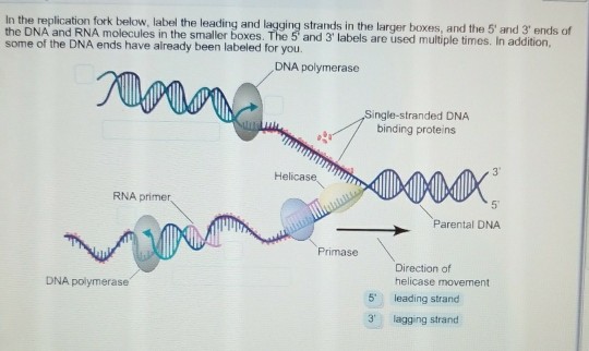
Solved In the replication fork below, label the leading and ...
DNA Replication Steps and Process - ThoughtCo Step 1: Replication Fork Formation. Before DNA can be replicated, the double stranded molecule must be "unzipped" into two single strands. DNA has four bases called adenine (A), thymine (T), cytosine (C) and guanine (G) that form pairs between the two strands. Adenine only pairs with thymine and cytosine only binds with guanine.

Draw a labelled diagram of replicating fork.
Solved Label the parts of the DNA replication fork. DNA - Chegg Question: Label the parts of the DNA replication fork. DNA helicase Okazaki fragment Open beta clamp DNA ligase DNA polymerase III Leading strand Closed beta clamp RNA primer DNA primase Single-strand binding proteins DNA gyrase Clamp loader DNA polymerase I Parent DNA This problem has been solved! See the answer Show transcribed image text

Chk1 promotes replication fork progression by controlling ...
PDF DNA Replication Drawing Name - AMAZING WORLD OF SCIENCE WITH MR. GREEN 1. On the diagram below, label the 5' and 3' ends of both parental DNA strands (you can make up which is which) 2. Label the replication fork 3. Draw and label helicase 4. Label the overall direction of DNA replication 5. Draw and label single stranded binding proteins 6. Draw and label the leading strand 7.

Schematic representation of different labelling variants as ...
Replication Fork: Definition, Structure, Diagram, & Function The replication fork is a structure which is formed during the process of DNA replication. It is activated by helicases, which helps in breaking the hydrogen bonds, and holds the two strands of the helix. The resulting structure has two branching's which is known as prongs, where each one is made up of single strand of DNA.

Label the figure below. Word bank: leading strand, lagging ...
Label DNA and Replication - Google Slides Label the diagram: DNA polymerase adds nucleotides (5' to 3') Replication fork is formed. DNA polymerase attaches to the primer. Okazaki fragments bound by ligase. DNA helicase unwinds DNA. Rearrange the steps to indicate the correct order: 1. Enzyme that unwinds DNA.
DNA Replication
Replication Fork | Science Primer The replication fork * is a region where a cell's DNA * double helix has been unwound and separated to create an area where DNA polymerases and the other enzymes involved can use each strand as a template to synthesize a new double helix. DNA Base RNA Base An enzyme called a helicase * catalyzes strand separation.

Solved Complete diagram of a replication fork in bacterial ...
Copy of DNA Replication - Labeling 1 - Name - StuDocu 63. Okazaki fragments Fragments of copied DNA created on the lagging strand. Primase Initiates the synthesis DNA by creating a short RNA segment at replication fork. Place the events in the correct order: Okazaki fragments are bound together by ligase 5 DNA helicase unwinds DNA biologycorner | Image Credit: Wikimedia Commons.

Replication Bubble Overview & Diagram | What is a Replication ...
DNA Replication Labeling Diagram | Quizlet An enzyme that creates a short RNA primer for initiation of DNA replication. RNA Primer short segment of RNA used to initiate synthesis of a new strand of DNA during replication The primer synthesized by primase enzyme DNA Polymerase on Leading Strand synthesizes new DNA only in the 5' to 3' direction Ligase

Draw a labelled schematic sketch of replication fork of DNA.
DNA Replication - Labeling worksheet with image - Name - StuDocu DNA Replication - Labeling (with word bank) DNA polymerase 3' 5' DNA Ligase Okazaki fragment DNA Primase ... ____ DNA polymerase adds nucleotides in the 5' to 3' direction ____ Replication fork is formed ____ DNA polymerase attaches to the primer ____ Okazaki fragments are bound together by ligase ____ DNA helicase unwinds DNA.

Free photo Label Diagram Dna Replication Labeled Biology ...
Draw a labelled diagram of replicating fork. - Toppr Ask DNA replicates in a semi-conservative manner in which each individual strand is copied to form a new molecule of DNA. The two strands can be labelled with isotopes using substrates that contain either normal 1 4 N or its heavy isotope 1 5 N.In an experiment, one strand of DNA was labelled with 1 4 N and the other with 1 5 N (hybrid DNA). The hybrid DNA was then allowed to replicate in the ...

DNA Replication Fork Quiz
A, schematic representation of the replication fork ...
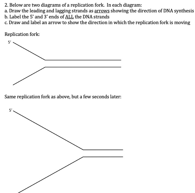
SOLVED:2. Below are two diagrams ofa replication fork In each ...

Replication Fork Diagram | Quizlet

DNA fibre labelling to measure replication fork rates and ...
this is a replication fork including one new but incomplete dna strand with the 5 end labeled_ direction of replication fork complete the diagram by label the box with an arrow to indicate t 39294
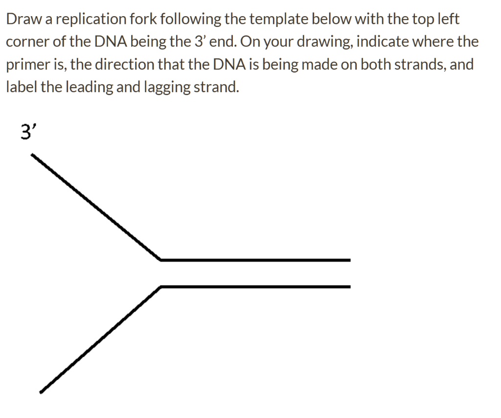
SOLVED:Draw a replication fork following the template below ...

Schematic of the experimental procedures used to identify ...
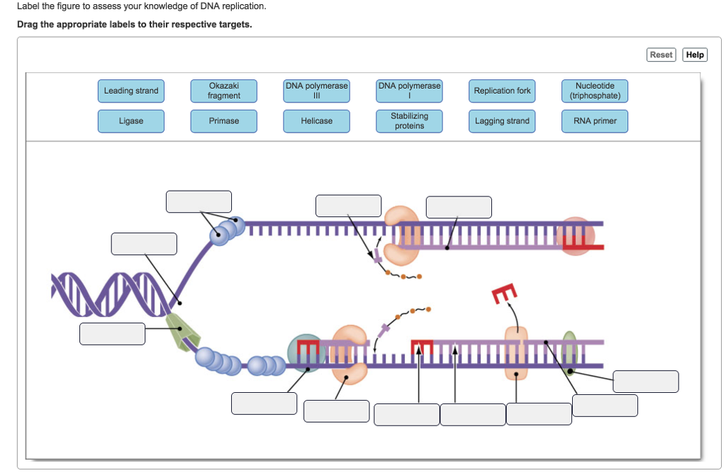
Answered: Label the figure to assess your… | bartleby
DNA Replication Replication Fork Enzyme Triangle PNG, Clipart ...
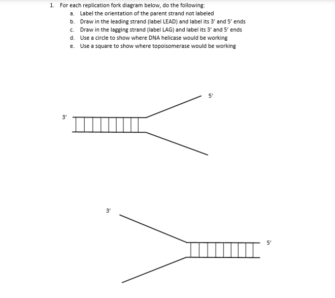
Solved a. 1. For each replication fork diagram below, do the ...

Micro 261 Exam 1- Chapter 4 Mastering Questions Flashcards ...
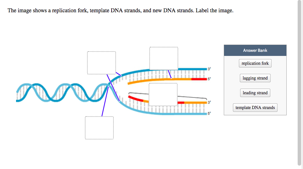
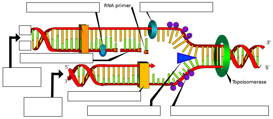
Solved The image shows a replication fork, template DNA ...
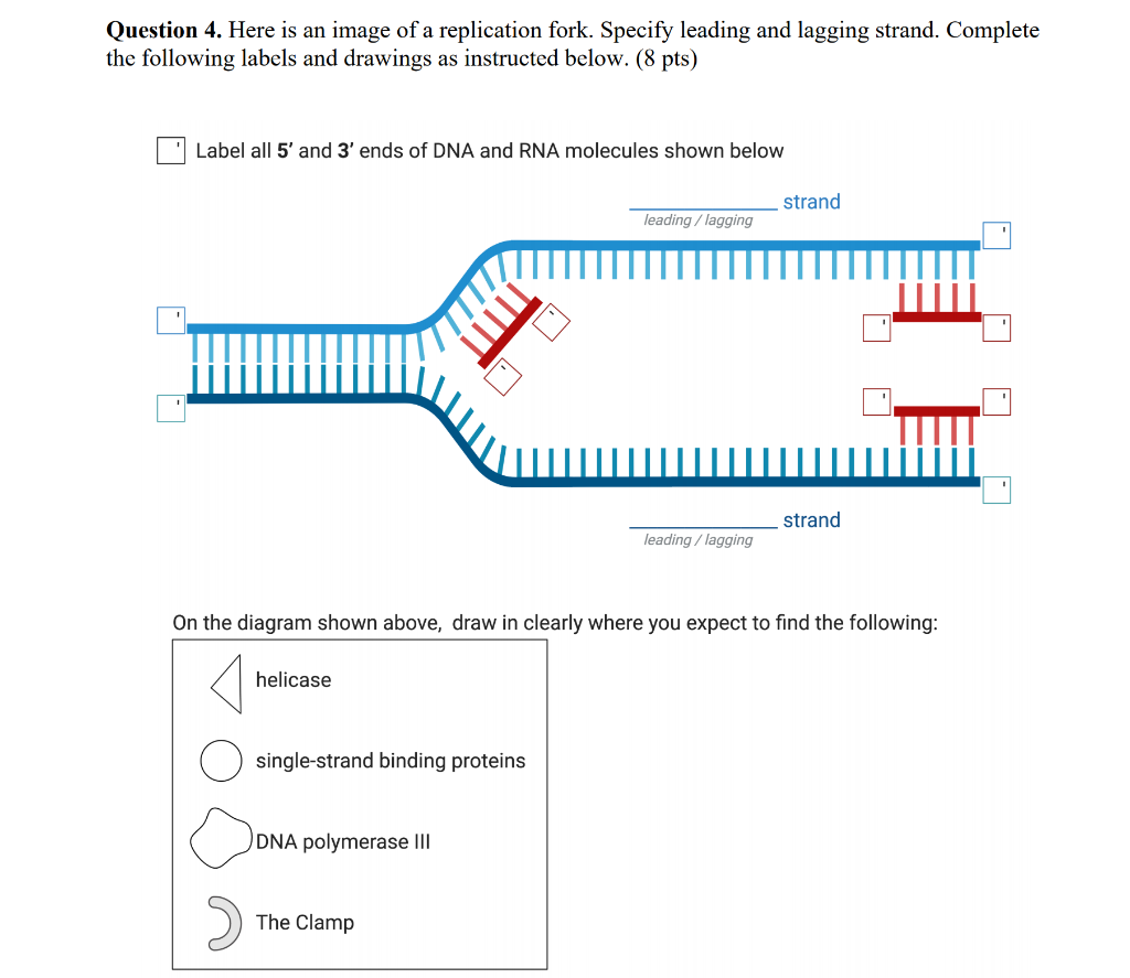
Solved Question 4. Here is an image of a replication fork ...
Molecular mechanism of DNA replication (article) | Khan Academy

7.1 DNA STRUCTURE AND REPLICATION HL
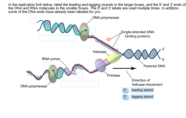
Solved In the replication fork below, label the leading and ...
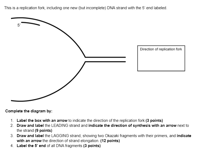
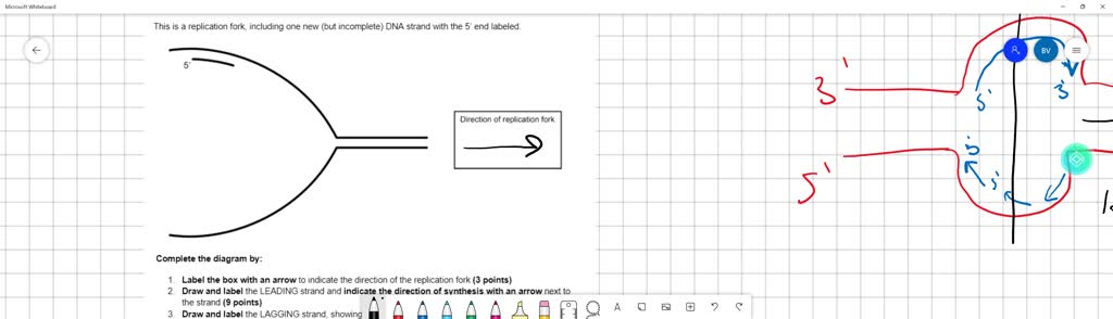
SOLVED:This is a replication fork, including one new (but ...

Scheme of the replication fork. a: template, b: leading ...

(a) Draw a labelled diagram of a \"replicating fork\" showing the polarity. Why does DNA replication

Lesson Explainer: DNA Replication | Nagwa

BIOEXCEL 190 - Molecular Genetics KEY

NAP1L1 and LANA were detected at replication forks on ...
Make a sketch of the double helix of DNA. Show how it unzips ...
Post a Comment for "38 label the replication fork"