38 label the photomicrograph
Label The Photomicrograph Of Thick Skin / Solved Label The ... Label The Photomicrograph Of Thick Skin / Solved Label The Photomicrograph Of Thin Skin Label The Photomicrograph 1 Answer Transtutors. Learn about dermatitis and how you can treat it Start studying photomicrograph of the epidermal layer in thick skin. Can you identify the five major layers of the epidermis? (Get Answer) - Label the photomicrograph in Figure 7.4. Examine a slide ... Label The Photomicrograph Of The Skin And Its Accessory Structures. Sebaceous Gland Duct Of Sebaceous Gland Epidermis Hair Follicle Posted 3 months ago. Q: Activity 4 Differentiating Sebaceous and Sweat Glands Microscopically Using the slide thin skin with hairs and the photomicrographs of cutaneous glands (Figure 7.6) as a guide, identify ...
Photomicrograph Atlas | U.S. Geological Survey - USGS.gov Apr 01, 2021 · The Photomicrograph Atlas provides a basic tutorial in the nomenclature of organic materials as they occur in sedimentary rocks such as coal and shale, information on the taxonomies used by various groups and organizations, and a database of images related to the characterization of fossil fuel resources in the United States and the world. Launch the Photomicrograph Atlas.

Label the photomicrograph
Ch. 22 Assessment Flashcards | Quizlet Label the structures in the photomicrograph based on the hints provided. List the correct order of lymphatic flow through a lymph node. 1. Afferent lymphatic vessel 2. Subcapsular sinus of the cortex 3. Sinuses of cortex and medulla 4. Efferent lymphatic vessel Put the following events into the correct order. 1. (Get Answer) - Label the photomicrograph based on the hints provided ... Label the photomicrograph based on the hints provided. Capsule Zona glomerulosa Zona fasciculata Capillaries Suprarenal gland Fascicle of cells Glomerulus of cells Mar 29 2022 11:26 AM [Solved] Please see an attachment for details | Course Hero Please see an attachment for details. Image transcription text. Label the photomicrograph based on the hints provided. Medulla Capillary Zona. fasciculara Suprarenal gland Zona reticularis... Show more. Biology Science Anatomy BISC 106.
Label the photomicrograph. Photomicrographs illustrating the different labels used to ... Photomicrographs illustrating the different labels used to identify new neurons in HVC at low (A-B) and high (C-D) magnifications. The first two panels show ... Solved Label the photomicrograph. Lumen Epithelial cell Transcribed image text: Label the photomicrograph. Lumen Epithelial cell Myoepithelial cell Apocrine sweat gland McGraw-Hil Education/Dennis Strete Label ... Label The Photomicrograph Of Thick Skin. - Exercise 4 Quiz Flashcards ... 1 answer to label the photomicrograph of thin skin. The epidermis, made of closely packed epithelial cells, and the dermis, made of dense, irregular connective tissue . Epidermis Of Thick Skin from eugraph.comThe skin is composed of two main layers: Thick skin showing epithelial detail. Practice labeling the layers of the skin. Label the photomicrograph in figure 128 figure 128 - Course Hero Label the photomicrograph in Figure 12.8Figure 12.8. LAB ACTIVITY 3: Neuromuscular Junction Examine a prepared microscope slide of the neuromuscular junction and identify the structures listed in Figure 12.8Figure 12.8.
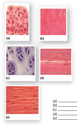
Label The Photomicrograph - Mr. Hill's Biology Blog: Our cells "inner ... Label the photomicrograph of thick skin. Use a label line and the letter p for each section. Monocyte, erythrocyte, lymphocyte, neutrophil, basophil, eosinophil. Schematically sketch and label the resulting microstructure. Place the following layers in order from superficial to deep. Solved Label the photomicrograph. Myoepithelial cell Lumen - Chegg Question: Label the photomicrograph. Myoepithelial cell Lumen Epithelial cell Apocrine sweat gland OMG Den Stree Myoepithelial cell Lumen Epithelial cell Apocrine sweat gland OMG Den Stree This problem has been solved! 1 2 label the photomicrograph of a transverse section 1 2 label the photomicrograph of a transverse section School Chamberlain College of Nursing Course Title ANATOMY 252 Type Lab Report Uploaded By sav001 Pages 20 Ratings 100% (41) This preview shows page 7 - 13 out of 20 pages. View full document See Page 1 Solved > Question 31 points Label the photomicrograph of thin:391984 ... 31 points Label the photomicrograph of thin skin. Hair Follicle Hair Dermis Sebaceous gland Duct of sebaceous gland Reset zoom. Solution. 5 (1 Ratings ) Solved. Biology 2 Years Ago 77 Views. This Question has Been Answered! View Solution. Related Answers.
Label The Photomicrograph Of Thick Skin : 6 6 Skin Photomicrographs Ta ... Label the photomicrograph of thick skin. Start studying photomicrograph of thick skin. Thick skin · stratum basale (also known as s. Part a is a micrograph showing a cross section of thin skin. (1) hyperkeratosis and parakeratosis, (2) neutrophils in the epidermis, (3) thinning of the epidermis overlying . Label the photomicrograph of thick skin. Endocrine Lab Worksheet Label the photomicrograph ... Question: Endocrine Lab Worksheet Label the photomicrograph based on the hints provided Capillary 0.25 points Exocrine portion Pancreas Print Pancreatic islet ... Final Exam A&P 1 Flashcards | Quizlet Label the photomicrograph of thin skin Hair shaft, epidermis, dermal root sheath, sebaceous gland, dermis, hair matrix label the structures of the hair follicle Identify the layers of the epidermis with relation to their location and role in keratinization ... the receptors responsible for olfaction are located in the olfactory epithelium The Flame's Daughter Ending Explained - Blogger Label the types of cells in the photomicrograph using the hints provided. PART A Structure of the Blood Vessel Wall 1. Identify each component of the electrical conduction system of the heart. Label the features of the head in midsagittal section. Label the photomicrograph in figure 74. Arteries have thicker walls than.
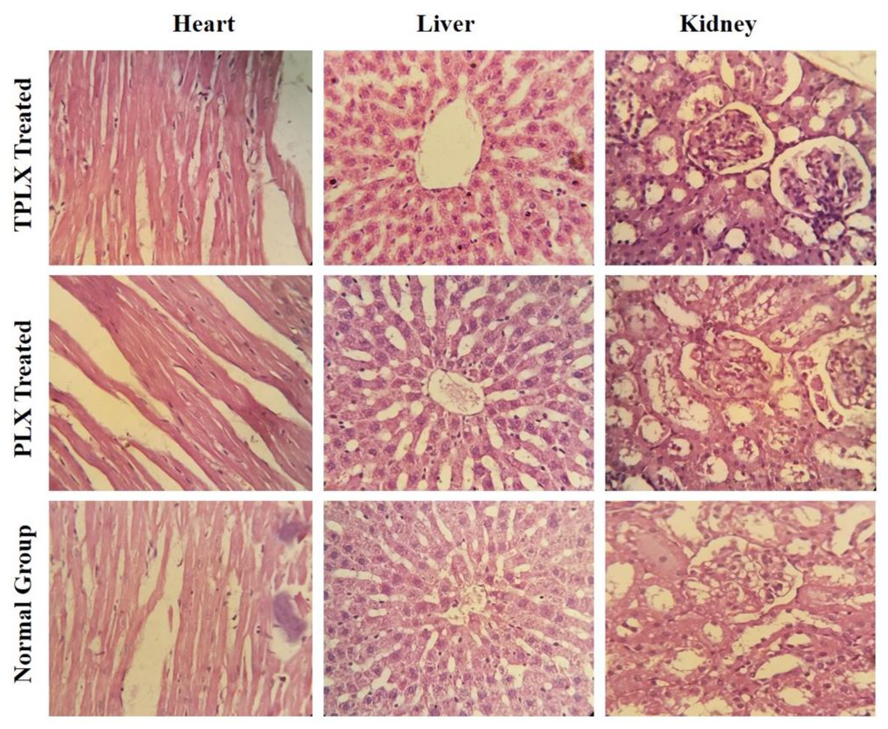
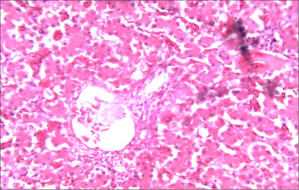
Photomicrograph of kidney sections of methotrexale treated ...
Label The Photomicrograph Of The Lung : 4 Chloro Dl Phenylalanine ... Label the photomicrogram of the lung segmental branch of pulmonary a. Make sure you know the basics of lung cancer, including prevention, risk factors, symptoms and treatment options. Label the anterior view of the lower respiratory tract based on the hints if. Electron micrograph of lung tissue (click to show / hide labels).
Solved Help Save & Exit Label the photomicrograph based on ...
Solved Label the photomicrograph based on the hints Question: Label the photomicrograph based on the hints provided. Capsule Zona glomerulosa Zona fasciculata Capillaries Suprarenal gland Fascicle of cells ...
Solved Lab 11 Endocrine System Saved Label the | Chegg.com
A & P lab test 4 Flashcards | Quizlet Label the photomicrograph based on the hints provided. Label the midsagittal view of the brain based on the hints provided. Which structure is highlighted? zona reticularis. zona fasciculata. Label the photomicrograph based on the hints provided. Label the histology of the ovary using the hints provided.

Photomicrograph of a transverse section in the jejunum of ...
Label The Photomicrograph Of Thick Skin - Faktor yang Start studying photomicrograph of thick skin. Apocrine sweat gland label the photomicrograph in figure 7.4. Learn vocabulary, terms, and more with flashcards, games, and other study tools. Label the photomicrograph of thick skin. 1 answer to label the photomicrograph of thin skin. The epidermis of thick skin has five layers:
Biomedicines | Free Full-Text | Intestinal Ischemia: Unusual ...
Label The Photomicrograph Of Compact Bone. / Tik Ta Lk Aitee Umoffong ... Solved Label The Photomicrograph Of Compact Bone Osteocyte Chegg Com from media.cheggcdn.com Label the photo micrograph of compact bone. Osteocyte central canal osteon canaliculus lacuna lamella central canal cement. Tyronecoleman332 is waiting for your help. Add your answer and earn points.

1,726 Immunofluorescent Photomicrograph Photos and Premium ...
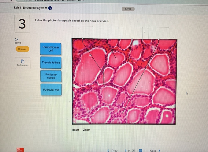
Endocrine System APR Module 8 Flashcards - Quizlet Label the photomicrograph based on the hints provided. (pancreas) Label the photomicrograph based on the hints provided. (suprarenal gland) Label each of the following histology slides by dragging the histology slide of the gland under the correct name.

1 2 Label the photomicrograph of a transverse section of the ...
BIOL 320 - Practical 2 - Lab 7 Flashcards - Quizlet Label the types of cells in the photomicrograph using the hints provided. Label the photomicrograph of the wall of the aorta using the hints provided. Label the photomicrograph of the wall of the inferior vena cava using the hints provided.

Endocrine Lab Flashcards | Quizlet
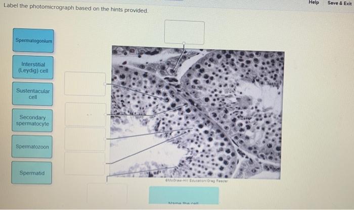
Label the Photomicrograph Using the Hints Provided. Drag each label into the appropriate position to identify what cell type is described by the label. Label the image of a compound light microscope using the terms provided. Label the testis and spermatic cord using the hints provided. Place the following pictures of white blood cells stained purple in the slides into the appropriate category.

Labeling of “starter” neural progenitor cells. (A ...
[Solved] Please see an attachment for details | Course Hero Please see an attachment for details. Image transcription text. Label the photomicrograph based on the hints provided. Medulla Capillary Zona. fasciculara Suprarenal gland Zona reticularis... Show more. Biology Science Anatomy BISC 106.
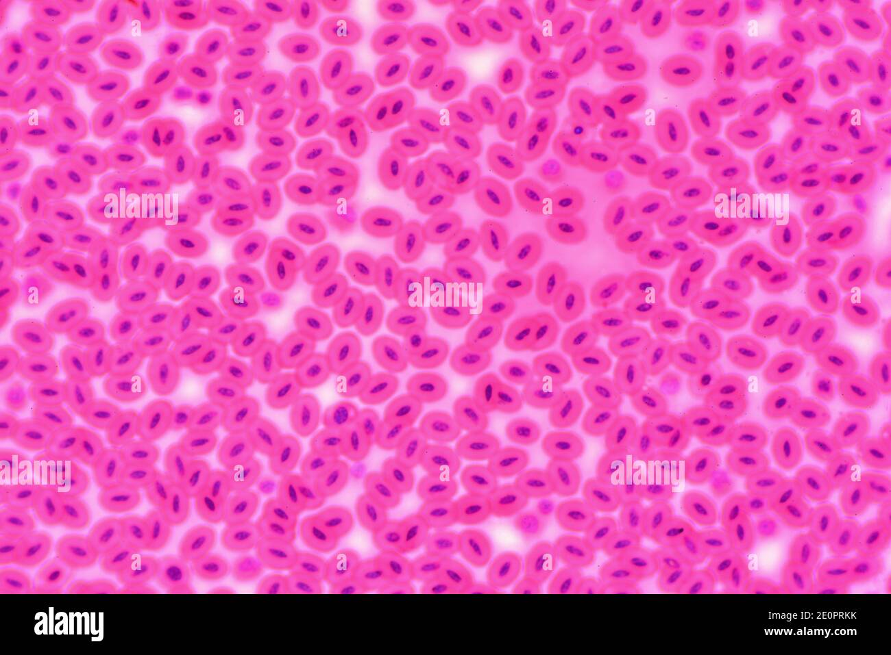
Snake blood. Red blood cells with nucleous. Photomicrograph ...
(Get Answer) - Label the photomicrograph based on the hints provided ... Label the photomicrograph based on the hints provided. Capsule Zona glomerulosa Zona fasciculata Capillaries Suprarenal gland Fascicle of cells Glomerulus of cells Mar 29 2022 11:26 AM

Light Photomicrograph Helianthus Stem Cross Section Stock ...
Ch. 22 Assessment Flashcards | Quizlet Label the structures in the photomicrograph based on the hints provided. List the correct order of lymphatic flow through a lymph node. 1. Afferent lymphatic vessel 2. Subcapsular sinus of the cortex 3. Sinuses of cortex and medulla 4. Efferent lymphatic vessel Put the following events into the correct order. 1.

3,644 Photomicrograph Stock Photos and Images - 123RF
Get Answer) - Label the following photomicrographs with the ...

Endocrine Lab Flashcards | Quizlet

Endocrine Lab Flashcards | Quizlet
Solved] Please see an attachment for details | Course Hero

Endocrine Lab Flashcards | Quizlet

5 The Integumentary System. - ppt download

hair - Search - Science Photo Library

4,730 Photomicrograph Photos - Free & Royalty-Free Stock ...
Pharmaceutics | Free Full-Text | Synthesis and Evaluation of ...

Photomicrograph (H and E stain, 40×) corresponds to Label 'B ...

Premium Photo | Photomicrograph of urine analysis showing ...

Cartilage in fetal finger. Photomicrograph Stock Photo - Alamy

Photomicrograph Of Simple Columnar Epithelium Of The ...
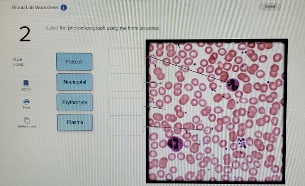
Solved Blood Lab Worksheet i Saved Label the photomicrograph ...

SciELO - Brasil - Ki67 Labelling Index predicts clinical ...

Gambar gratis: tebal, darah smear, photomicrograph, koma ...

The MEMIC: An ex vivo system to model the complexity of the ...

Protection effects of rice protein hydrolysate on UVB ...

Cell Mitosis-photomicrograph art print poster

Photomicrograph showing myoepithelial cells | Download ...
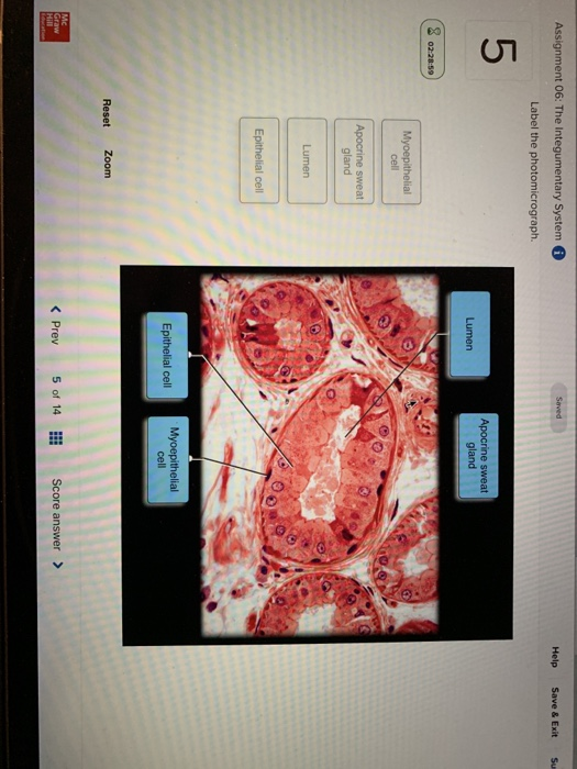
Solved Assignment 06: The Integumentary System Help Save ...

Liposarcoma Human Photomicrograph Panorama Seen Under Stock ...
Therapeutic Evaluation of Neemazal® Against Experimental ...
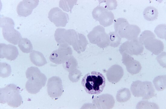
Gambar gratis: darah smear, plasmodium malariae ...

Light Photomicrograph Cucurbita Stem Cross Section Stock ...
Portofolio Foto dan Gambar Stok dari Jubal Harshaw | Shutterstock
Post a Comment for "38 label the photomicrograph"