44 label the skin structure and areas indicated
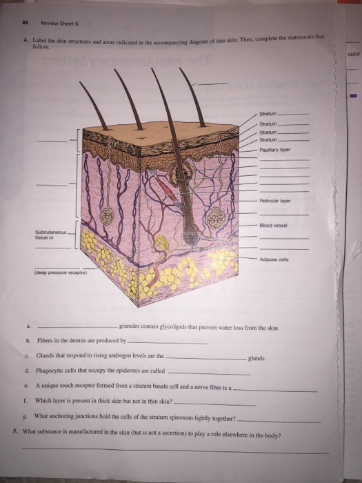
Solved 4. Label the skin structures and areas indicated in ... 4. Label the skin structures and areas indicated in the accompanying diagram of thin skin. Then, complete the that follow Stratum Stratum Stratum. Stratum Popilary layer Reticular layer Blood vessel tissue or Adpose cels (deep pressure receptor granules contain glycolipids that prevent water loss from the skin. b. Fibers in the dermis are ... PDF Label The Skin Structures And Areas Indicated Label The Skin Structures And Areas Indicated Penrose tiling Wikipedia. Therapeutic Injections for Pain Management Types of. All ecolabels Ecolabel Index. ... April 28th, 2018 - Treatment of all skin and skin structure problems including conditions of the hair nails and mucous membranes including the mouth and genital regions''WorkSafeBC
Skin Structure (Labeling) Flashcards - Quizlet Start studying Skin Structure (Labeling). Learn vocabulary, terms, and more with flashcards, games, and other study tools.
Label the skin structure and areas indicated
Solved 4. Label the integumentary structures and areas | Chegg.com Label the integumentary structures and areas indicated by leader lines in the figure below. AS 5. Label the layers of the epidermis in thick skin. Then, complete the statements that follow. a. Glands that respond to rising androgen levels are the glands. . are epidermal cells that play a role in the immune response. PDF Label The Skin Structures And Areas Indicated integumentary system basic structure of the skin, review sheet 7 the integumentary system name lab time, student objectives lake county schools overview, the lumbar plexus spinal nerves branches teachmeanatomy, hazard symbol wikipedia, solved label the skin structures and areas indicated in, skin structure diagram to label anatomy structure ... 5.2 Accessory Structures of the Skin - Anatomy & Physiology Chapter Review. Accessory structures of the skin include hair, nails, sweat glands, and sebaceous glands. Hair is made of dead keratinized cells, and gets its color from melanin pigments. Nails, also made of dead keratinized cells, protect the extremities of our fingers and toes from mechanical damage. Sweat glands and sebaceous glands produce ...
Label the skin structure and areas indicated. PDF Label The Skin Structures And Areas Indicated Label The Skin Structures And Areas Indicated Dermagen Skin Care Fusion Labs. Chapter 2 Definitions California Fire Code 2016 UpCodes. ATA 100 Chapters s techent com. Chapter 64 Agriculture and Natural Resources Based. Carbofuran C12H15NO3 PubChem. Federal Register Bar Code Label Requirement for Human. Accessory Structures of the Skin - Anatomy and Physiology Hair. Hair is a keratinous filament growing out of the epidermis. It is primarily made of dead, keratinized cells. Strands of hair originate in an epidermal penetration of the dermis called the hair follicle.The hair shaft is the part of the hair not anchored to the follicle, and much of this is exposed at the skin's surface. The rest of the hair, which is anchored in the follicle, lies ... Structure of Skin - Explore its Parts and Function - BYJUS It is a fleshy surface with hair, nerves, glands and nails. It consists of hair follicles which anchor hair strands into the skin. It acts as a barrier between outside and inside environment. The skin has different thicknesses and textures. E.g. the skin under the eyes is as thin as paper but is thick at the soles of the feet and palm. MODULE 4.docx - Lesson 1: 28 Label the skin structures and areas ... EPIDERMIS The epidermis is the skin's outermost layer, made up of epithelial tissue. This layer of skin acts as a barrier and is the first line of defense against viruses. 9. DERMIS The dermis, positioned between the epidermis and subcutaneous tissues, also known as the subcutis, is the second and thickest of the three primary layers of skin. 10.
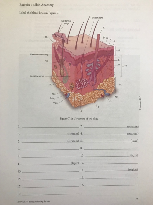
PDF wedgwood science Label the skin structures and areas indicated by leader lines and brackets on the figure. Select different colors for the structures below and color the coding cir- cles and the corresponding structures on the figure. Arrector pili muscle (D Adipose tissue C) Hair follicle C) Nerve fibers (C) Sweat (sudoriferous) gland Sebaceous gland Figure 4—2 8. PDF Label The Skin Structures And Areas Indicated wikipedia, skin structure diagram to label anatomy structure, 7 2 the skull anatomy and physiology opentextbc ca, 4 label the skm structures and areas indicated in the, anatomy and physiology of animals the skin wikibooks, the lumbar plexus spinal nerves branches teachmeanatomy, powerpoint presentation, printables skin diagram to PDF Label The Skin Structures And Areas Indicated wiley, skin structure and function definition diagrams, name period 4 skin and body membranes, 4 label the skm structures and areas indicated in the, the skin boundless anatomy and physiologypart 1 orientation and guide to the anatomy of the pelvis and perineum a self instructional module you should Assignment 11 pg 104.pdf - 4. Label the skin structures and ... 4. Label the skin structures and areas indicated in the accompanying diagramofthin skin. Then, complete the statements thatfollow. SubcutaneousJtissue or _l T~P-r
PDF Label The Skin Structures And Areas Indicated april 30th, 2018 - 149 azalea circle • limerick pa 19468 1330 610 495 6898 office • 484 680 0507 cell e mail s tech''skin cancer treatment pdq® —health professional version january 31st, 2018 - expert reviewed information summary about the treatment of skin cancer''federal register bar code label requirement for human 2-1 Mastering A&P Lab Module Two Homework - Part A - Skin Structures ... Without skin, humans would quickly fall prey to infections and perish from water and heat loss. Part A - Major Layers of Skin The skin comprises three distinct layers--the epidermis, dermis, and hypodermis. Each is composed of different tissue types. Match the appropriate layer of skin to its description. Hint 1. Consider tissue type ... PDF Label The Skin Structures And Areas Indicated 'name lab time date review sheet the integumentary system april 30th, 2018 - 4 label the skin structures and areas indicated in the accompanying diagram of thin skin then complete the statements that follow a granules extruded from the keratinocytes prevent water loss by diffusion through''dermagen skin care fusion labs april 26th, 2018 - love … 5.1 Layers of the Skin - Anatomy & Physiology The hypodermis (also called the subcutaneous layer or superficial fascia) is a layer directly below the dermis and serves to connect the skin to the underlying fascia (fibrous tissue) surrounding the muscles. It is not strictly a part of the skin, although the border between the hypodermis and dermis can be difficult to distinguish.
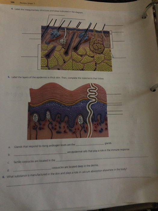
Solved: 100 Sheet 7 4. Label The Integumentary Structures ... | Chegg.com
PDF Label The Skin Structures And Areas Indicated Label The Skin Structures And Areas Indicated red colored skin may also occur as a result of blood vessels in or near the skin dilating expanding due to ... skin structure diagram to label is free hd wallpaper this wallpaper was upload at march 10 2017 upload by admin in structure

Conceptual Marketing Corporation - ANALYSIS INFORMATION FROM A EUROPEAN ...
PDF Label The Skin Structures And Areas Indicated Label The Skin Structures And Areas Indicated ... care skin cancer surgery read about signs of skin cancer if skin will return what to do the day of surgery and what to do after surgery''federal register bar code label requirement for human february 25th, 2004 - information about this document as published in the federal register this tables of ...

30 Label The Skin Structures And Areas Indicated In The Accompanying ...
Layers of the Skin - Anatomy and Physiology The skin is composed of two main layers: the epidermis, made of closely packed epithelial cells, and the dermis, made of dense, irregular connective tissue that houses blood vessels, hair follicles, sweat glands, and other structures. Beneath the dermis lies the hypodermis, which is composed mainly of loose connective and fatty tissues.

Conceptual Marketing Corporation - ANALYSIS INFORMATION FROM A EUROPEAN ...
PDF Label The Skin Structures And Areas Indicated Label The Skin Structures And Areas Indicated AR 40 5 PREVENTIVE MEDICINE OCCUPATIONAL SAFETY AND. CARBOFURAN C12H15NO3 PUBCHEM. WOUND CARE MEDICAL CLINICAL POLICY BULLETINS AETNA. DERMAGEN SKIN CARE FUSION LABS. PENROSE TILING WIKIPEDIA. SKIN CANCER TREATMENT PDQ® —HEALTH PROFESSIONAL VERSION. LABEL MOTOMCO.
33 Label The Skin Structures - Labels Database 2020
PDF Label The Skin Structures And Areas Indicated Dermagen Skin Care Fusion Labs. Wound Care Medical Clinical Policy Bulletins Aetna. Dermagen Skin Care Fusion Labs. Mohs Laser Surgery Amp Dermatopathology Surgeons And. Wound Care Medical Clinical Policy Bulletins Aetna. AR 40 5 Preventive Medicine Occupational Safety And. General Dermatology Dermatology And Allergy Specialists. All Ecolabels ...

34 Integumentary System Diagram To Label - Labels Design Ideas 2020
100 Review Sheet 7 4. Label the integumentary structures and areas ... 100 Review Sheet 7 4. Label the integumentary structures and areas indicated in the diagram. 5. Label the layers of the epidermis in thick skin. Then, complete the statements that follow EUR a. Glands that respond to rising androgen levels are the glands are epidermal cells that play a role in the immune response.

Conceptual Marketing Corporation - ANALYSIS INFORMATION FROM A EUROPEAN ...
Solved tive tissue 4. Label the skin structures and areas - Chegg Label the skin structures and areas indicated in the accompanying diagram of thin skin. Then, complete the statements that follow. Weisshaft Stratum opidamist Stratum Stratum Stratum Papilary layer Dermis Reticular layer ascectors allmusde Encrine Sweet blond Dermal Vascular plexus pensery neare fiber Blood vessel Subcutaneous tissue or.

PDF The Integumentary System - Holly H. Nash-Rule, PhD Label the skin structures and areas indicated in the accompanying diagram of thin skin. Then, complete the statements that follow. a. Lamellar granules contain glycolipids that prevent water loss from the skin. b. Fibers in the dermis are produced by fibroblasts . c.
31 Label Of The Skin - Labels Information List
Answers.docx - LESSON 1 STRUCTURE OF THE SKIN A. Label the skin ... LESSON 1 STRUCTURE OF THE SKIN A.Label the skin structures and areas indicated by the leader lines and brackets on the figure. Select different colors for the structures below. Describe the structure and functions of each layer. Epidermis-is the outer and thinner layer of the skin composed of keratinized stratified squamous epithelium.
Post a Comment for "44 label the skin structure and areas indicated"