39 anterior view of the heart with labels
Pink Floyd - Atom Heart Mother | Releases | Discogs referencing Atom Heart Mother (LP, Album, Limited Edition, Numbered, Reissue, Remastered) MFSL 1-202 On my copy the vinyl itself weighs 203.5 grams. Agree with the previous reviewer that the cover photos have this saturated, overexposed quality that diminishes a lot of fine detail, and MoFi wasn't using the heavier cardboard at this point. Coronary artery dominance - wikidoc Co-Dominance. Shown below is an image depicting co-dominant coronary artery. A coronary artery is said to have a "co-dominance" or balanced dominance when only the right posterior descending artery (RD or RPDA) arises from the right coronary artery (RCA), while the circumflex inferior artery (CI) and the circumflex posterior artery (CP) arise from the circumflex artery (CX).
Spinal cord injury - Wikipedia A person with a mild, incomplete injury at the T5 vertebra will have a much better chance of using his or her legs than a person with a severe, complete injury at exactly the same place. Of the incomplete SCI syndromes, Brown-Séquard and central cord syndromes have the best prognosis for recovery and anterior cord syndrome has the worst.

Anterior view of the heart with labels
1: Anatomy of the Heart (Anterior View): The figure illustrates the ... Atrial fibrillation (AF) is the most common cardiac arrhythmia in United States. The most popular treatment for AF is a percutaneous procedure called catheter ablation. Current AF ablation... Chapter 19: The Heart Flashcards | Quizlet -Heart muscle receives blood when the ventricles relax •Left coronary artery (LCA)-anterior interventricular branch •supplies blood to interventricular septum and anterior walls of ventricles-circumflex branch •passes around left side of heart in coronary sulcus, supplies left atrium and posterior wall of left ventricle Label the Heart Diagram Anterior view Quiz - PurposeGames.com This is an online quiz called Label the Heart Diagram Anterior view There is a printable worksheet available for download here so you can take the quiz with pen and paper. Your Skills & Rank Total Points 0 Get started! Today's Rank -- 0 Today 's Points One of us! Game Points 24 You need to get 100% to score the 24 points available Actions 2 favs
Anterior view of the heart with labels. Circulatory System Coloring and Labeling - Lovejoy Anatomy and ... - Google SUPERIOR ASPECT OF THE HEART This view of the heart is seen as if the atria and the major vessels have been removed. You should be able to see all of the major valves of the heart. The most anterior valve is the pulmonary semilunar valve that occurs between the right ventricle and the pulmonary trunk. Label and color this valve blue.Posterior to this is the aortic semilunar valve. Heart Anatomy: Labeled Diagram, Structures, Blood Flow ... - EZmed There are 3 tricks to remember the tricuspid valve is located on the right side of the heart, and the mitral valve is located on the left. Trick 1: You can use the saying "TRI (Try) it before you BI (buy) it". This will help you remember the tricuspid valve comes before the bicuspid/mitral valve. Chapter 22 Heart Flashcards - Quizlet Label the coronary arteries in an anterior view of the heart. Label the order that blood flows through in the heart, using the arrows as guides. Label the components of the heart wall. Label the components of the heart as seen from a posterior view. Label the major coronary veins. Label the components of the conduction system. Heart Anatomy: Heart Dissection - University of Washington The picture below shows an anterior view of the heart with the pericardium removed. The letters indicated in the text refer to the labels on the picture. The anterior surface of the heart is characterized by the presence of the large arteries leaving the base of the heart, the pulmonary trunk (H) and the aorta (G).
Heart Labeling anterior view Diagram - Quizlet Heart Labeling anterior view STUDY Learn Write Test PLAY Match + − Created by Meghan12th PLUS Terms in this set (26) brachiocephalic trunk ... left common carotid artery ... superior vena cava ... aortic arch ... liigamentum arteriosum ... right pulmonary artery ... amending aorta ... right pulmonary veins ... pulmonary trunk ... right atrium ... Heart Anatomy - Posterior View - Maricopa Heart Anatomy: Posterior View When you point to any structure on the photograph, that region or structure will be highlighted in the smaller image to the left to help you locate it. ... Posterior Heart Tutorial and Self-Test to review basic anatomy of heart and vessels... Quizzer 1 on basic EKG features including deflections and segments etc ... Anatomy Tutorial - Anterior | Atlas of Human Cardiac Anatomy This illustration demonstrates an anterior view of the thoracic cavity, highlighting the position of the heart in relationship to the ribs and diaphragm. The right atrium, right ventricle, and a small portion of the left ventricle are visible from this aspect. Note that in the majority of cases, 2/3 of the heart is positioned to the left of ... Heart Anatomy - Anterior (Front) View : Medical Illustration This medical exhibit pictures an anterior (front) view of the heart anatomy with labels for the aorta, superior vena cava, right atrium, right ventricle, inferior vena cava, pulmonary trunk, left atrium, pulmonary veins and left ventricle. Max Image Size: 2448 pixels wide by 1804 pixels high. Recent Comments. No comments have been posted.
Lab 44- Heart Structure Flashcards - Quizlet Label the anterior heart structures by clicking and dragging the labels to the correct location. 1. Aoric Arch 2.Super vena cava ... 10.Right ventricle 11. Inferior vena Cava (Straight down.) Label the internal heart structures frontal section (anterior view) by clicking and dragging the labels to the correct location. On the left: (6) 1.)Right ... The Heart - Science Quiz - GeoGuessr The Heart - Science Quiz: Day after day, your heart beats about 100,000 times, pumping 2,000 gallons of blood through 60,000 miles of blood vessels. If one of your organs is working that hard, it makes sense to learn about how it functions! This science quiz game will help you identify the parts of the human heart with ease. Blood comes in through veins and exists via arteries—to control the ... Anterior View Human Body Illustrations & Vectors Rib Cage of Human Skeleton System Anatomy with detailed labels Anterior View. 3D Illustration Concept of Rib Cage of Human Skeleton System Anatomy with detailed. Human body front, back and side views. ... The human heart anatomy Anterior View, Frontal section and cardiac muscle structure. Spine Vertebrae - Lateral view / Side view. Human body ... External Anatomy of the Heart- Anterior View - Maricopa Anterior View of Sheep Heart: Point to any region of the large image, that region will then be highlighted in the smaller image to the left to help you locate it. If you click your left mouse button, the name of that region will appear to identify it. ... Quizzer 2 on EKG arrhythmias such as heart blocks, PAC, digitalis effect etc...
Anomalous origin of the right coronary artery from the left anterior ...
Solved Heart Chambers and Valves: Frontal Heart Section 2 - Chegg Heart Chambers and Valves: Frontal Heart Section 2 Label the chambers and valves seen in an anterior view of the heart Left ventricle Right ventricle Aortic valve Tricuspid Valve Right atrum Mihral valve Chord Pulmonary ave

cardiac anatomy and physiology revision :: www.forensicmed.co.uk
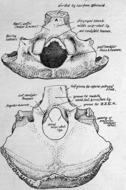
Radiological anatomy of the spine - e-Anatomy - IMAIOS Sep 13, 2021 · Standard radiographic view of anatomical structures of the spinal column. On "Anatomical parts" the user can choose between three types of labels: vertebrae, bones and joints. On "Series" the user can select the radiographs concerning the spine as a whole, the cervical, thoracic and lumbar vertebrae, the sacrum and coccyx.

Muscle Flashcards Flashcards by ProProfs
Blausen 0451 - Anterior view of the heart - English labels Anterior view of the heart. English labels. By Blausen Medical Communications, Inc. Retrieved from Wikimedia Commons: category: Images from Blausen Medical Communications. Fig. 0451. Anatomical structures in item: Cor. Auricula dextra. Auricula sinistra. Truncus pulmonalis. Arcus aortae.

Anterior interventricular coronary artery - The Anatomy of… | Flickr
Solved Label the structures seen in the anterior view of the - Chegg View the full answer Transcribed image text: Label the structures seen in the anterior view of the heart. Superior vena cava Interior vena cava Aorta Left atrium Pulmonary trunk Pulmonary vein Left ventricle Pulmonary artery Right ventricle Right atrium Previous question Next question

MBBS Medicine (Humanity First): Heart Murmurs
anterior heart Quiz - PurposeGames.com This is an online quiz called anterior heart. ... This quiz has tags. Click on the tags below to find other quizzes on the same subject. quiz. heart. label. anterior. Your Skills & Rank. Total Points. 0. Get started! Today's Rank ... Add to tournament 1 tournaments. Printables and Stats. View as Printable Worksheet. Game Statistics. Give a nod ...

Cross Sections Through the Thorax
Heart Diagram with Labels and Detailed Explanation - BYJUS Well-Labelled Diagram of Heart. The heart is made up of four chambers: The upper two chambers of the heart are called auricles. The lower two chambers of the heart are called ventricles. The heart wall is made up of three layers: The outer layer of the heart wall is called epicardium. The middle layer of the heart wall is called myocardium.

BIO 211 (UNIT # 1) Lab Practicum flashcards | Quizlet
Posterior heart view and labels Diagram | Quizlet bring oxygen-rich blood from the left lung to the left atrium Auricle of left atrium paired, only portion of left atrium seen in anterior view Left atrium Chamber that receives oxygenated blood from the pulmonary veins and pumps it into systemic circulation. Great cardiac vein runs alongside the anterior interventricular artery Coronary sinus
11 Best Images of Horse Anatomy Blank Worksheet - Sacrum and Coccyx ...
Human Heart - Diagram and Anatomy of the Heart - Innerbody The heart is a muscular organ about the size of a closed fist that functions as the body's circulatory pump. It takes in deoxygenated blood through the veins and delivers it to the lungs for oxygenation before pumping it into the various arteries (which provide oxygen and nutrients to body tissues by transporting the blood throughout the body).
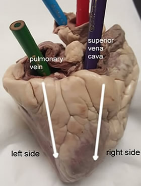
Sheep Heart Dissection
Whitesnake (album) - Wikipedia Initially the album was released worldwide with different titles, tracklists and by different record labels. In Europe and Australia, it was titled 1987 and included two extra songs absent from the North American version, "Looking for Love" and "You're Gonna Break My Heart Again", while in Japan the album was released as Serpens Albus with the ...
Post a Comment for "39 anterior view of the heart with labels"