40 sketch and label a single motor neuron
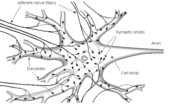
Draw a labelled diagram of the neuron and describe the ... - Toppr Ask The structure of neuron: Nerve cells or neurons are the structural and functional units of the nervous system. It consists of three major parts namely, Cell body, dendrites, Axon. Cell Body: It is irregular in shape or polyhedral. The cell body contains cytoplasm and certain granular bodies called Nissles granules which contain a group of ... Neuromuscular junction: Parts, structure and steps - Kenhub Each individual muscle fiber is innervated (supplied) and controlled by a motor neuron. This motor neuron, which has its cell body located within the central nervous system, will have axons that enter the muscle and penetrate the perimysium. At this point, each axon of the motor neuron will divide into branches called axon terminals.
MOTOR UNITS AND MUSCLE TWITCHES - Brigham Young University-Idaho One alpha motor neuron along with all of the muscle fibers it innervates is a motor unit . The size of the motor unit correlates with the function of the muscle. In muscles involved with fine, coordinated control, the motor units are very small with 3-5 muscle fibers per motor neuron.
Sketch and label a single motor neuron
Nervous Tissues and Nerves Flashcards | Quizlet Motor neuron Transmits impulse out of the brain or spinal cord to effectors Oligodendrocyte Myelin-forming neuroglial cell in brain and spinal cord Sensory neuron Transmit impulse into brain or spinal cord from receptors Single motor neuron Single sensory neuron Single neuroglial cell Single nerve fiber with schwann cell (cross-section) Motor Neuron: Function, Types, and Structure | Simply Psychology Motor neurons are responsible for integrating signals from the brain to the muscles, glands, and organs that intend to carry out the required motor function. Motor neurons allow us to move, talk, eat, swallow, and breathe, therefore without these cells, we would not be able to complete many basic life functions. Label Parts of a Neuron Diagram - Quizlet Only $35.99/year Label Parts of a Neuron STUDY Learn Flashcards Write Spell Test PLAY Match Gravity Created by cottonje Terms in this set (14) Dendrites receives impulses from other nerve cells axon hillock The cell body...the part of the cell that houses the nucleus and keeps the entire cell alive and functioning Myelin Sheath
Sketch and label a single motor neuron. Neuron Structure and Classification - Google Search Students can sketch and label the structure of a typical neuron, and describe the functions of each component. Students can classify neurons on the basis of their structure and function. Check... germanydating.expatica.comExpat Dating in Germany - chatting and dating - Front page DE The first and the best free dating site for Expats in Germany. Find and meet other expats in Germany. Register for free now. Motor Neuron - The Definitive Guide | Biology Dictionary A motor neuron is a cell of the central nervous system. Motor neurons transmit signals to muscle cells or glands to control their functional output. When these cells are damaged in some way, motor neuron disease can arise. This is characterized by muscle wasting ( atrophy) and loss of motor function. Motor Neuron Overview How to draw a Motor Neuron - YouTube This channel is about Education, Diagram drawing, Tour & Travel, DIY ideas, Mehndi designs, Gifting ideas, Origami ideas, Embroidery work, Card making, Poster making and drawing videos like border ...
Labeled Neuron Diagram - Science Trends Motor neurons are part of the central nervous system (CNS) and communicate signals from the spinal cord to the parts of the body to control their motion. For example, motor neurons send signals to the muscles in your arms causing them to contract. Motor neurons send electrical signals to your intestines so they move and churn food. journals.lww.com › neurosurgeryNeurosurgery - LWW Neurosurgery Speaks! Neurosurgery is proud to offer audio abstracts in 11 different languages, translated and read by native speakers. Each is the scientific abstract from a published article. Neuromuscular Junction Structure and Functions - New Health Advisor The synapse or connection between a motor neuron and a skeletal muscle is known as neuromuscular junction. Communication happens between the neuron and muscle via nerve cells. Due to this communication or transmission of signal, the muscle is able to contract or relax. It is the most widely studied synapse and it is comparatively easier to ... Peripheral nerves: Histology and clinical aspects | Kenhub A single motor neuron may innervate several to several hundred muscle cells; collectively the unit is referred to as a motor end plate. The telodendria are rich in synaptic vesicles and mitochondria. The synaptic vesicles contain acetylcholine (ACh) , which is released from the telodendria under the influence of the action potential.
10.2 Skeletal Muscle - Anatomy & Physiology (a) It is the number of skeletal muscle fibers supplied by a single motor neuron. (b) A large motor unit has one neuron supplying many skeletal muscle fibers for gross movements, like the Temporalis muscle, where 1000 fibers are supplied by one neuron. A small motor has one neuron supplying few skeletal muscle fibers for very fine movements ... PDF Nervous System Review Answer Key 2014 - Mayfield City Schools Created Date: 3/14/2014 1:02:16 PM Neuron Structure and Classification - Google Search Students can sketch and label the structure of a typical neuron, and describe the functions of each component. ... Checkpoints. Draw and label the structure of a neuron using the terms listed in the notes and book. ... Motor neurons - a nerve cell forming part of pathway along which impulses pass from the brain or spinal cord to a muscle or ... Neurons (With Diagram) - Biology Discussion It is a motor nerve and carries impulses from the brain to these muscles for controlling the movements of eyeball. IV. Trochlear Nerve: This nerve's name means "pulley" because it innervates an extrinsic eye muscle that loops a pulley-shaped ligament in the orbit. The trochlear nerve is the thinnest and smallest cranial nerve.
Schwann Cell Anatomy - Human Anatomy - GUWS Medical Sketch and label a single motor neuron in the space provided in Part C of the laboratory report. 7. Obtain a prepared microscope slide of a dorsal root ganglion. Search the slide and locate a cluster of sensory neuron cell bodies. You also may note bundles of nerve fibers passing among groups of neuron cell bodies (fig. 25.4). 8.

Motor Neuron Diagram Labeled Sketch Coloring Page
Label Parts of a Neuron Diagram - Quizlet Only $35.99/year Label Parts of a Neuron STUDY Learn Flashcards Write Spell Test PLAY Match Gravity Created by cottonje Terms in this set (14) Dendrites receives impulses from other nerve cells axon hillock The cell body...the part of the cell that houses the nucleus and keeps the entire cell alive and functioning Myelin Sheath

Motor Neuron: Function, Types, and Structure | Simply Psychology Motor neurons are responsible for integrating signals from the brain to the muscles, glands, and organs that intend to carry out the required motor function. Motor neurons allow us to move, talk, eat, swallow, and breathe, therefore without these cells, we would not be able to complete many basic life functions.
Diagram Flower Black White Vector Stock Vector 109995023 - Shutterstock
Nervous Tissues and Nerves Flashcards | Quizlet Motor neuron Transmits impulse out of the brain or spinal cord to effectors Oligodendrocyte Myelin-forming neuroglial cell in brain and spinal cord Sensory neuron Transmit impulse into brain or spinal cord from receptors Single motor neuron Single sensory neuron Single neuroglial cell Single nerve fiber with schwann cell (cross-section)

2. Nerve and Muscle Cells
Post a Comment for "40 sketch and label a single motor neuron"